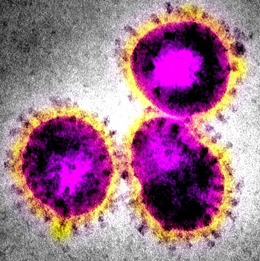
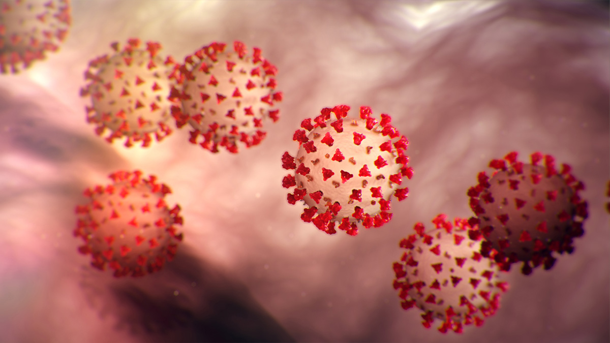
15 + Coronavirus Image Under A Microscope High Quality Images. Timelapse video shot at Melbourne's Doherty Institute for Infection and Immunity shows a sample of the coronavirus successfully growing in the laboratory. This isn't quite as sharp as the first one, but you can see the spikes on the surface Viruses in the coronavirus family only have small differences in their genome, with only five nucleotide differences between three of the viruses.

21 + Coronavirus Image Under A Microscope High Quality Images
The discovery by researchers at Westlake University in.
Pin en High Resolution Photo
Coronavirus: Images of cells from scanning electron ...
LOOK: Under The Microscope, Deadly Viruses Look Absolutely ...
Here’s the Coronavirus Under an Electron Microscope
ZOOM : BEGINILAH COVID-19 : CORONAVIRUS UNDER MICROSCOPE ...
USA confirms 1st Wuhan coronavirus case
ELI5: Do some viruses really look like "robots"? If so ...
Coronavirus Utah: Department of Health announces second ...
Coronavirus - What is it, what are the risks and what do ...
Pin on Microbiology class (Bio253) board
Top stories: A push to build affordable electron ...
Coronavirus Under Microscope 5 Unbelievable Facts About ...
‘Preparedness, not containment' for COVID-19 | MUSC ...

Adjusting to coronavirus threat | United Methodist News ...
Never-before-seen virus may be behind mystery outbreak in ...
15 + Coronavirus Image Under A Microscope Desktop WallpaperEnglish: Coronaviruses are a group of viruses that have a halo, or crown-like (corona) appearance when viewed under an electron microscope. An electron micrograph of a thin section of MERS-CoV. This is due to the presence of viral spike peplomers emanating from each proteinaceous envelope.

