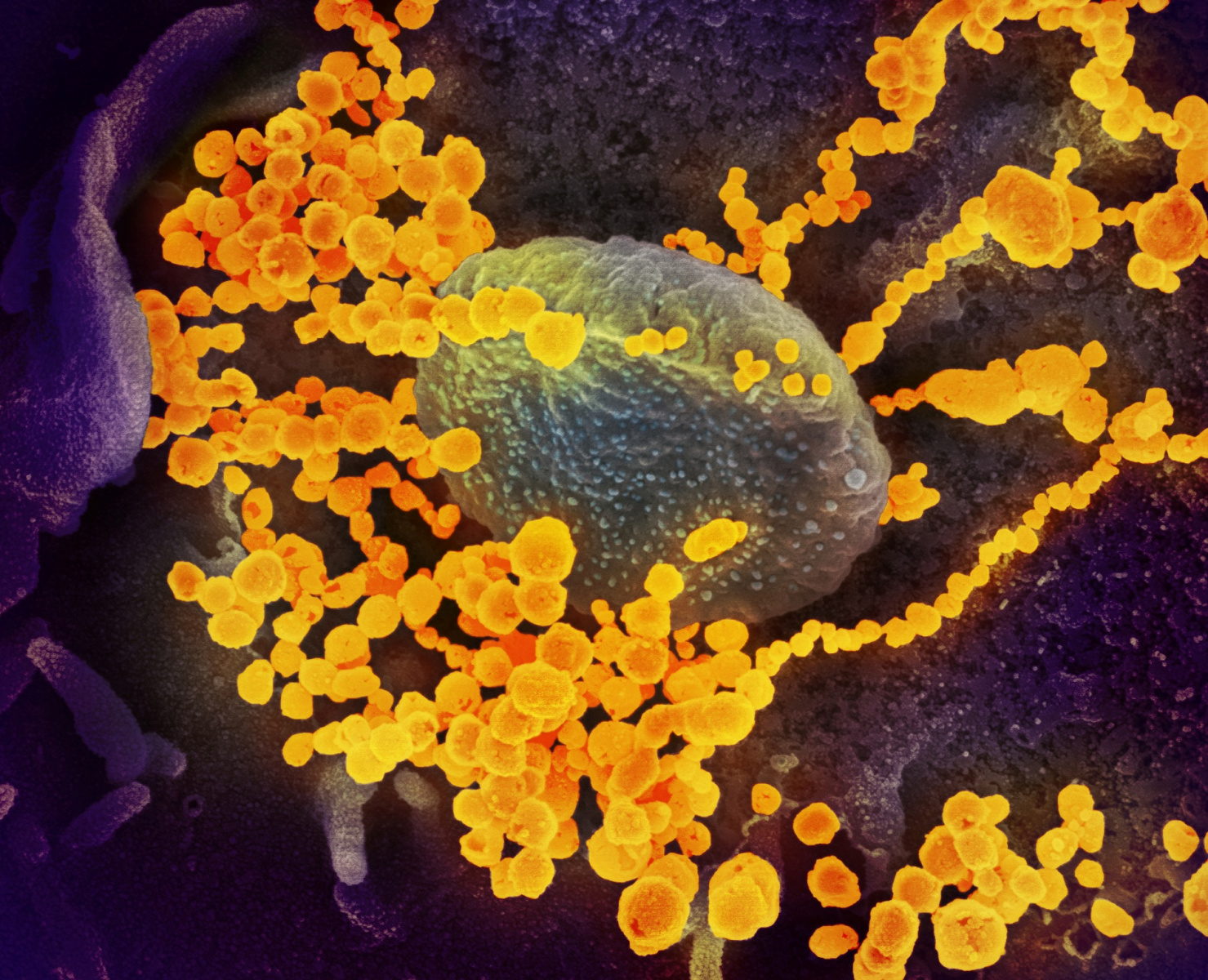
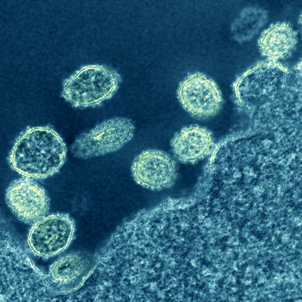
15 + Coronavirus Real Image Microscope Background Images. Thousands of new, high-quality pictures added every day. This scanning electron microscope image shows the new coronavirus (yellow) among human cells (blue, pink and purple).

21 + Coronavirus Real Image Microscope Background Images
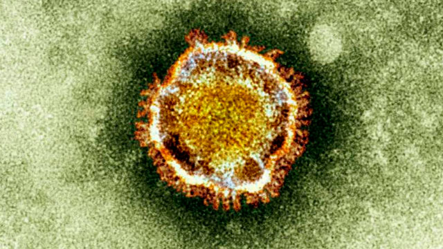
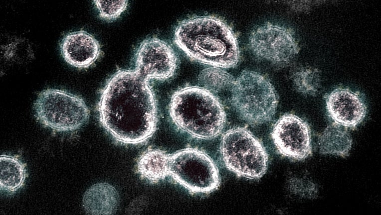
If these look a little familiar, it's because most coronaviruses - such as SARS and MERS - look similar on the outside, sharing the bump-covered spherical appearance.
UPDATED: Two Central Va. patients test negative for ...


NSW researchers breakthrough on the novel coronavirus ...
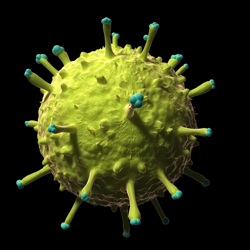
These 12 Viruses Look Beautiful Up Close But Would Kill ...

Coronavirus | New Scientist

Coronavirus kills off BRAIN cells, study finds - Angle News

Laboratorydeal - Corona Virus Microscope – laboratorydeal

COVID-19 | Schultz Realty
Ibuprofen and COVID-19 symptoms – here’s what you need to know
SARS-Like Virus Vaccine Unlikely, Experts Say - ABC News

MD Coronavirus Totals: 12,830 Confirmed Cases, 486 Deaths ...
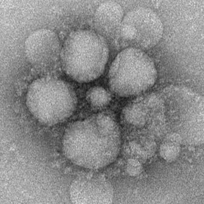
1918 H1N1 Virus Particles | Electron micrograph of 1918 ...

As Novel Coronavirus Outbreak Continues, WHO, CDC Urge ...
Santa Clara County reports 59 new COVID-19 cases, total at ...
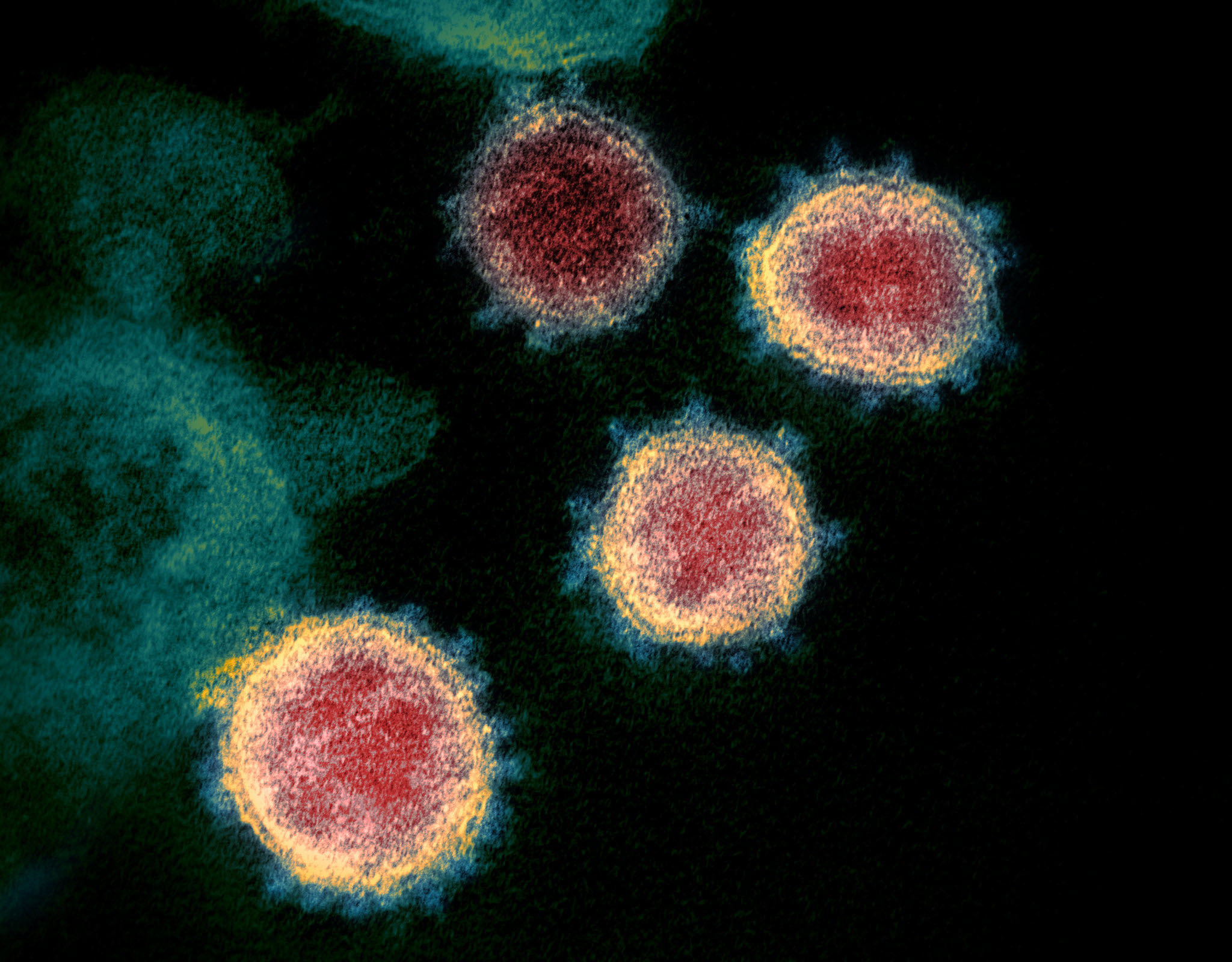
Novel coronavirus structure reveals targets for vaccines ...

Coronavirus - Wikipedia
15 + Coronavirus Real Image Microscope HD ResolutionsS., emerging from the surface of cells A laboratory in the United States has produced the most detailed images to date of the novel coronavirus currently spreading across the globe. They were made with scanning and transmission electron microscopes. Jupyter Notebook tutorials on solving real-world problems with Machine Learning & Deep Learning using PyTorch.