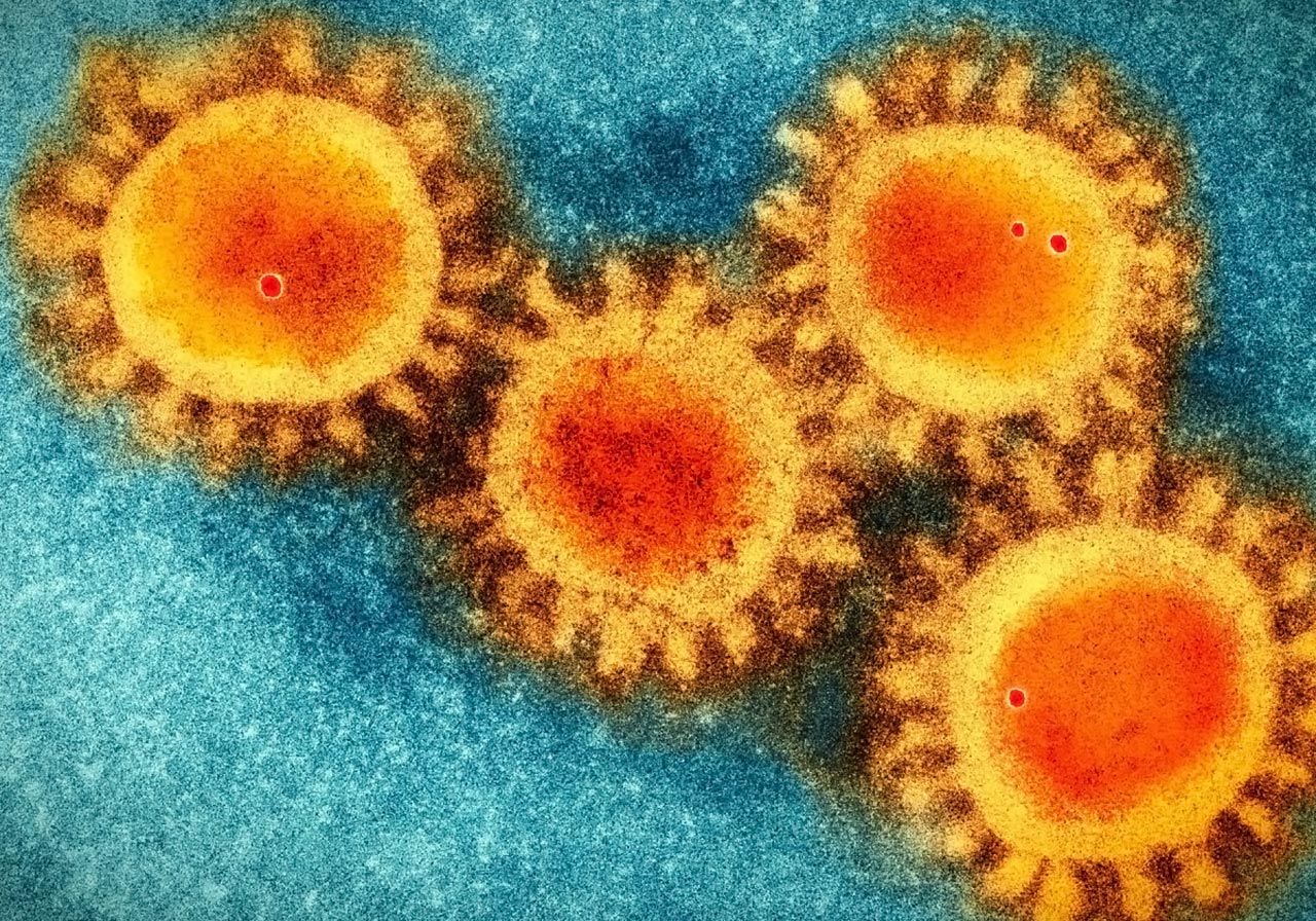
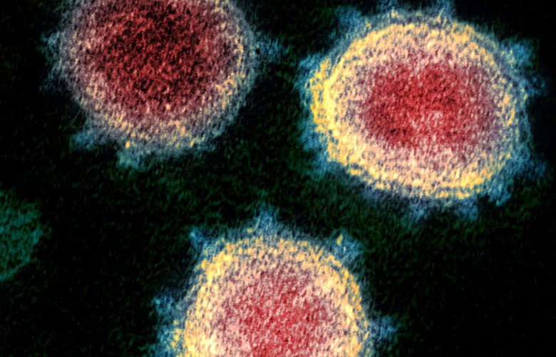
15 + Electron Microscope Images Of Coronavirus High Quality Images. This isn't quite as sharp as the first one, but you can see the spikes on the surface Viruses in the coronavirus family only have small differences in their genome, with only five nucleotide differences between three of the viruses. This is due to the presence of viral spike peplomers emanating from each proteinaceous envelope.

21 + Electron Microscope Images Of Coronavirus HD Wallpapers
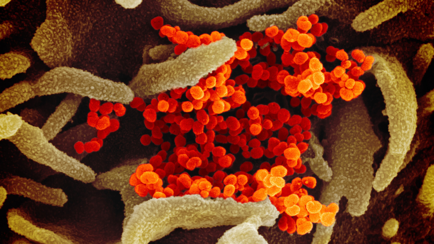
The 'invisible' enemy unmasked: Chilling microscope images reveal the reality of coronavirus as it erupts out from the surface of a human cell.
No Evidence COVID-19 Coronavirus Was Genetically ...


Severe acute respiratory syndrome coronavirus - Wikipedia

Senator asks gig companies to fight coronavirus by helping ...

Coronavirus electron-microscope look - Kostenlose ...

SARS virus particles Electron microscopy of numerous SARS ...
Adjusting to coronavirus threat | United Methodist News ...

IMAGES: What New Coronavirus Looks Like Under The ...

4th U.S. Coronavirus Case Confirmed In Los Angeles County

Picture of a coronavirus as seen through an electron ...

There Are Now 8 Presumptive Positive Coronavirus Cases In ...
HSS response to COVID-19 coronavirus: patients and visitors
$2M fund started to help gig workers, vulnerable ...

Opinion | These Coronavirus Exposures Might Be the Most ...
Could COVID-19 Spur A Revolution In Vaccine Development?
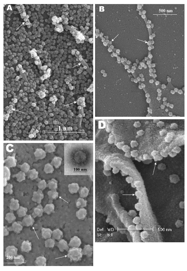
Figure 3 - Topographic Changes in SARS Coronavirus ...
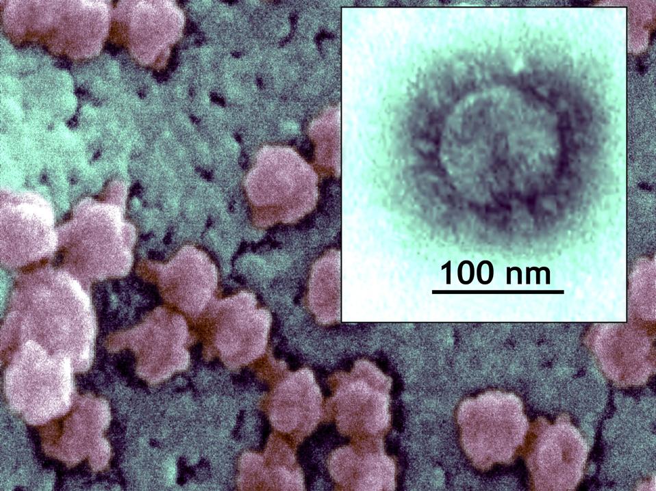
15 + Electron Microscope Images Of Coronavirus HD ResolutionsThis scanning electron microscope image shows the new coronavirus (yellow) among human cells (blue, pink and purple). Pune: The first images from India of what the coronavirus looks like are out. The virus is seen emerging from the surface of cells cultured in a lab and isolated from a patient in the US.