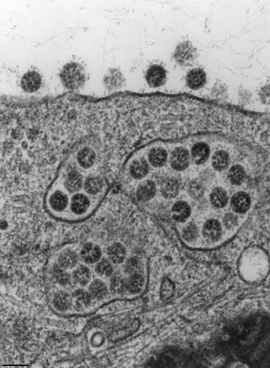
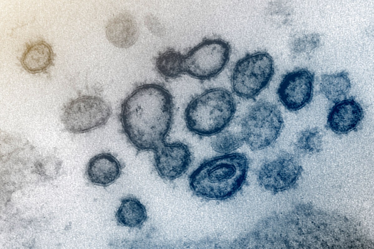
15 + Electron Microscope Image Of Covid 19 Background Images. Negative stain electron microscopy shows a MERS-CoV particle with club-shaped surface projections surrounding the periphery of the particle, a characteristic feature of coronaviruses. To better delineate the virus from healthy.
21 + Electron Microscope Image Of Covid 19 HD Wallpapers
One such test screens patient blood samples for antibodies against the virus.
.jpg)
2019 coronavirus virus officially named COVID-19


IMAGES: What New Coronavirus Looks Like Under The ...

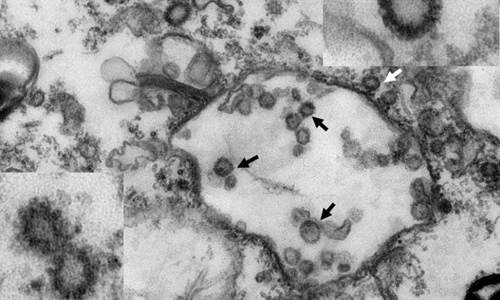
Figure. Electron microscopy images of thin sections and ...
WHO officially names the novel coronavirus as ‘COVID-19 ...
Here’s the Coronavirus Under an Electron Microscope

UKMALAYALEE | HOME

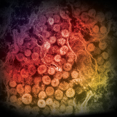
Picture of a coronavirus as seen through an electron ...

Coronavirus Testing Is Available But Not Everyone Needs to ...
Major U.S. stock indexes drop for third straight day
‘Preparedness, not containment' for COVID-19 | MUSC ...


Murrieta high school closed, 71 students self-quarantined ...
The why’s behind COVID-19 survival and immunity ...

Top stories: A push to build affordable electron ...

Pin on Electron microscopy

There Are Now 8 Presumptive Positive Coronavirus Cases In ...
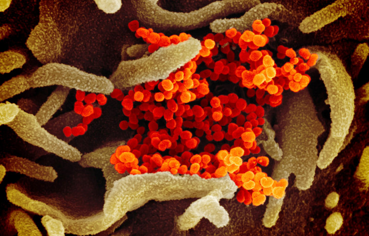
15 + Electron Microscope Image Of Covid 19 Background ImagesTaken by scanning electron microscope, images were colourised to better delineate virus from healthy cells. Negative stain electron microscopy shows a MERS-CoV particle with club-shaped surface projections surrounding the periphery of the particle, a characteristic feature of coronaviruses. The Electron Microscope (EM) is an impressively powerful microscope available today, allowing researchers to view a specimen at nanometer size.